All content on this site is intended for healthcare professionals only. By acknowledging this message and accessing the information on this website you are confirming that you are a healthcare professional. If you are a patient or carer, please visit the International Myeloma Foundation or HealthTree for Multiple Myeloma.
The Multiple Myeloma Hub website uses a third-party service provided by Google that dynamically translates web content. Translations are machine generated, so may not be an exact or complete translation, and the Multiple Myeloma Hub cannot guarantee the accuracy of translated content. The Multiple Myeloma Hub and its employees will not be liable for any direct, indirect, or consequential damages (even if foreseeable) resulting from use of the Google Translate feature. For further support with Google Translate, visit Google Translate Help.


The Multiple Myeloma Hub is an independent medical education platform, sponsored by Bristol Myers Squibb, GSK, Johnson & Johnson Innovative Medicine, Legend Biotech, Pfizer, Roche, and Caribou Biosciences. Funders are allowed no direct influence on our content. The levels of sponsorship listed are reflective of the amount of funding given. View funders.
Now you can support HCPs in making informed decisions for their patients
Your contribution helps us continuously deliver expertly curated content to HCPs worldwide. You will also have the opportunity to make a content suggestion for consideration and receive updates on the impact contributions are making to our content.
Find out more
Create an account to access:
Bookmark & personalize site content
Receive alerts for new content in your areas of interest
View multiple myeloma content recommended for you
Multiple myeloma: An overview
Do you know... How many times more likely is multiple myeloma in male versus female patients?
Multiple myeloma (MM) is a monoclonal gammopathy, resulting from the abnormal proliferation of clonal plasma cells. This abnormality leads to an excess production of a single immunoglobulin and, in turn, high levels of monoclonal paraprotein M – also referred to as the M protein. MM is the second most common hematological malignancy diagnosed in adults, with the highest prevalence among older male patients from non-White backgrounds.1
Etiology
The primary cause of MM is not fully understood, but it is believed to be the result of an acquired genetic mutation. Translocations in chromosome 14 are commonly observed in MM, and common oncogene mutations and their incidence in MM include.2
- KRAS: 36%
- NRAS: 20%
- TP53: 16%
- DIS3: 16%
- FAM46C: 12%
The main risk factors associated with the development of MM include3
- Older age (≥65 years)
- Male gender
- Men are 1.5 times more likely to develop MM than women
- Race
- Black patients are at a higher risk of developing MM compared with White patients
- A family history of MM
- Obesity
- Diagnosis of other plasma cell dyscrasias
- Monoclonal gammopathy of undetermined significance
- Solitary plasmacytoma
- Smoldering MM
Epidemiology
The age-standardized rate (ASR) of MM globally is 1.78 people per 100,000, with the highest incidence recorded in Australia and New Zealand (ASR: 4.86) and the lowest in western Africa (ASR: 0.81).4
Across all regions, MM is most prevalent in males, particularly in those over 65 years5; see Figure 1 for more details.
Figure 1. Epidemiology of multiple myeloma*
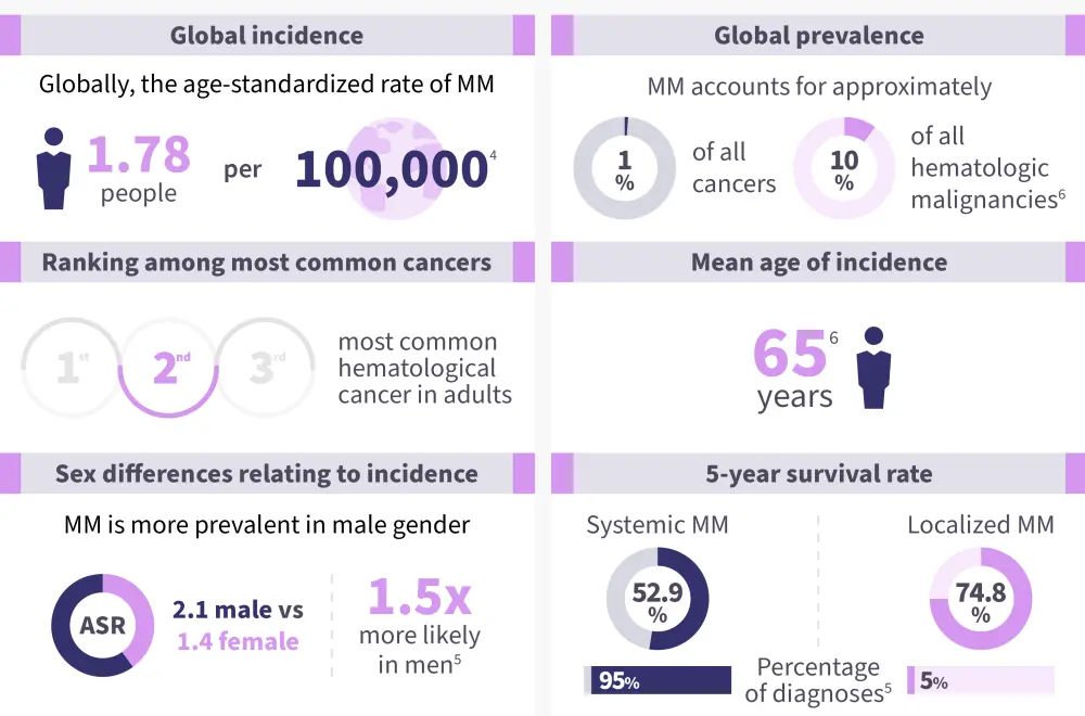
*Data from Huang, et al.4 and Padala, et al.5
Pathophysiology7,8
The pathophysiology of multiple myeloma is demonstrated in Figure 2.
Figure 2. Pathophysiology of multiple myeloma*
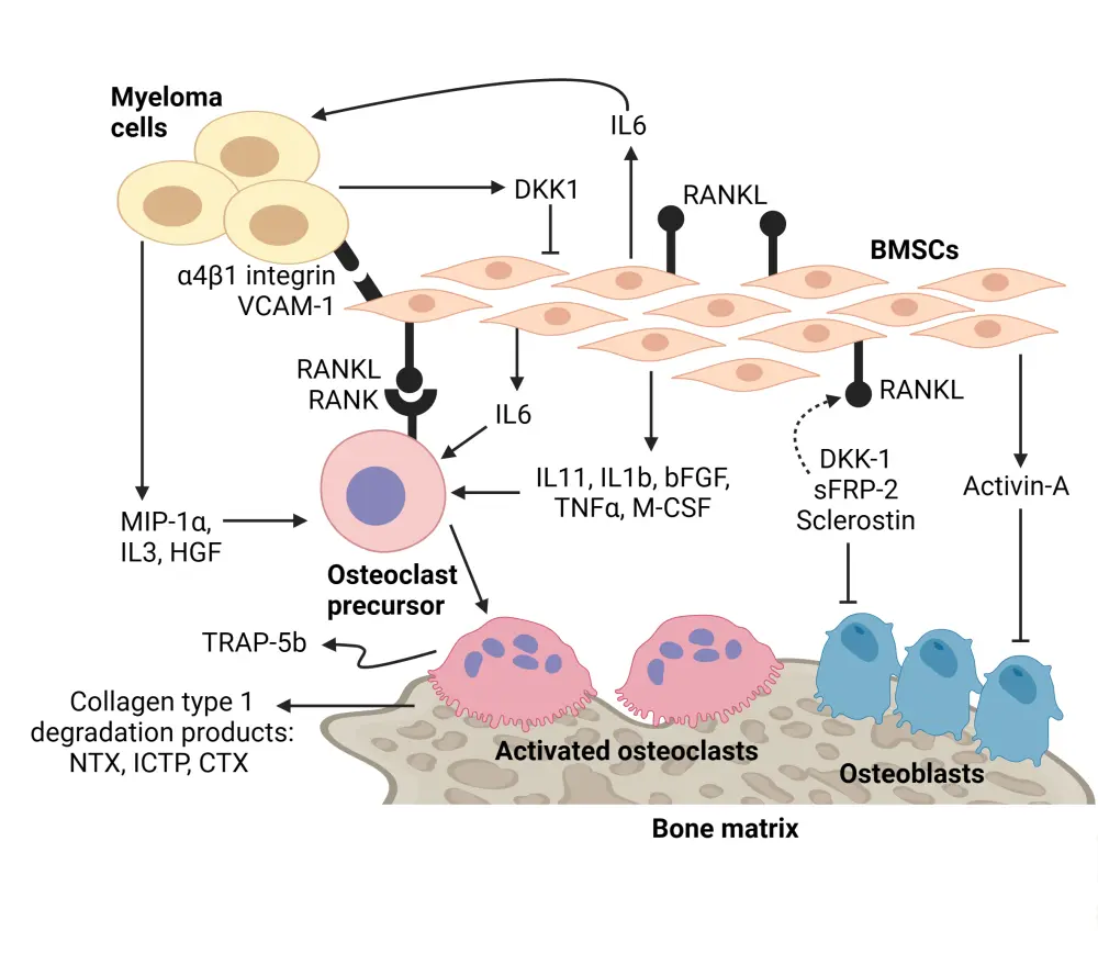
BMSC, bone marrow-derived mesenchymal stem cell.
*Adapted from Lentzsch, et al.7 and Mukkamalla, et al.8
Created with BioRender.com.
- Hematopoietic stem cells in the bone marrow differentiate into a disproportionate volume of B cells and, subsequently, overproduce plasma cells (>10% plasma cells).
- These plasma cells produce abnormal antibodies and an excess of light-chain antibodies.
- MM cells secrete cytokines such as IL3, which inhibits the production of osteoblasts, limiting the production of bone cells.
- MM cells cause an increase in osteoclast production and subsequent breakdown of bone cells by:
-
- Secreting DKK1, which inhibits OPG production by osteoblasts and, in turn, increases osteoclast activity
- Stimulating osteoclasts through the expression of MIP1α and RANKL
- Stimulating IL6, which is involved in the self-regulation of osteoclast cells.
- An increase in osteoclast activity results in the breakdown of bone cells, which then release calcium, and causes hypercalcemia.
- The paraproteins produced in excess cause renal damage and failure.
- Anemia can result from renal failure and the overproduction of plasma cells.
Signs and symptoms1
The primary indicators for MM are often denoted using the acronym CRAB:
C: hypercalcemia
R: renal failure
A: anemia
B: lytic bone lesions
Other common signs and symptoms are shown in Figure 3.
Figure 3. Signs and symptoms of multiple myeloma*
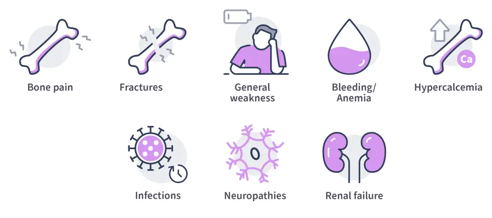
*Data from Van de Donk, et al.1
Diagnosis6
A diagnosis of MM requires at least one myeloma-defining event (MDE) to occur in addition to either ≥10% clonal plasma cells or an observed plasmacytoma.
MDEs may include any of the CRAB signs (listed above), as well as:
- Clonal plasma cells ≥60%
- Serum free light-chain ratio ≥100
- ≥1 focal bone lesion.
The high abundance of the M protein associated with MM can be used as a diagnostic indicator through serum protein electrophoresis. Further investigation with immunofixation can be used to determine MM subtype by identifying the particular immunoglobulin in excess.
Disease staging can then be determined using a number of tools, most commonly the Revised International Staging System (R-ISS), as shown in Table 1.
Table 1. Revised International Staging System for multiple myeloma*
|
CA, cytogenetic abnormalities; iFISH, interphase fluorescence in situ hybridization; LDH, lactate dehydrogenase; R-ISS, Revised International Staging System. |
|
|
Stage |
Criteria |
|---|---|
|
I |
|
|
II |
|
|
III |
and either
or
|
Risk stratification in MM is determined by the presence of following indicators for high-risk disease status:
- High risk genetic abnormalities:
- t(4;14)
- t(14;16)
- t(14;20)
- Del 17p or p53 mutation
- Gain 1q (amplification of duplication)
- High-risk plasma cell S-phase
- R-ISS stage 3
However, it is important to note that the individual criteria for diagnosis, risk stratification, and staging may vary by region. A variety of international guidelines can be found at the end of this document.
Management6,9
The only potentially curative treatment for MM is stem cell transplantation; thus, the MM treatment paradigm is differentiated by transplant eligibility. A summary of the current treatment paradigm is shown in Figure 4.
Figure 4. The multiple myeloma treatment paradigm*
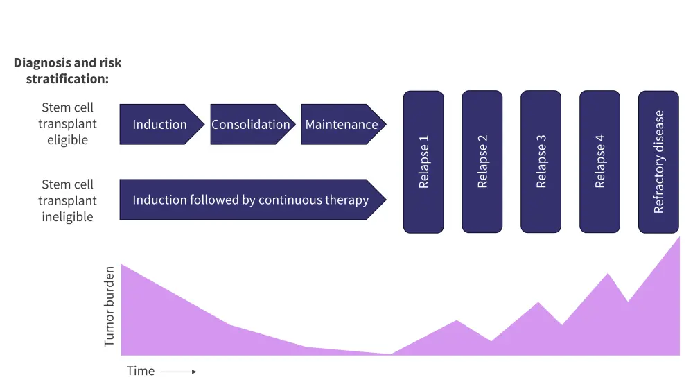
*Adapted from Rajkumar S.6
The recommendations for therapies in newly diagnosed MM, differentiated by transplant eligibility and risk status, are outlined in Figure 5.
Figure 5. Treatment recommendations for A transplant eligible and B transplant ineligible newly diagnosed multiple myeloma*
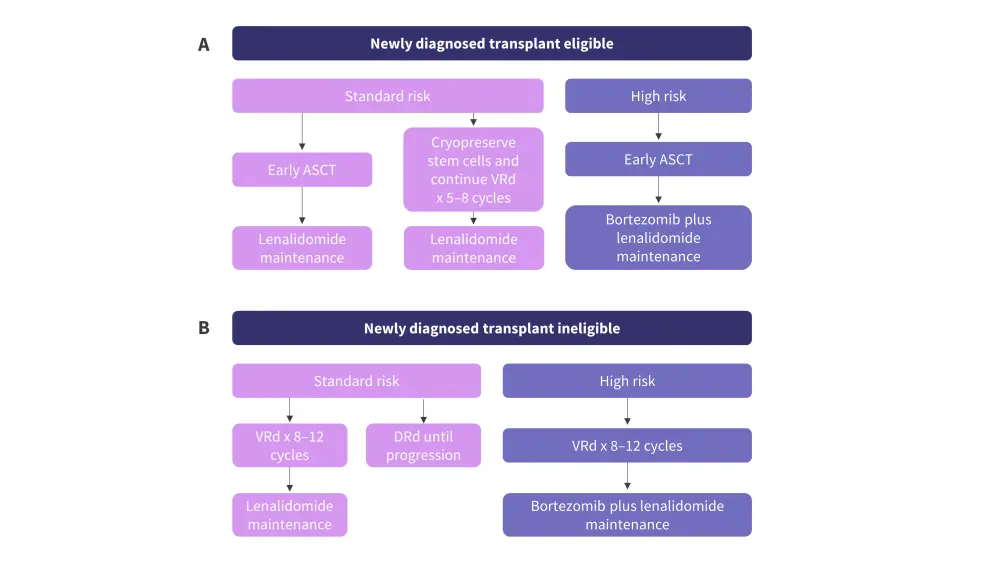
ASCT, autologous stem cell transplant; DRd, daratumumab lenalidomide dexamethasone; VRd, bortezomib lenalidomide dexamethasone.
*Adapted from Rajkumar S.6
The treatment of relapsed/refractory MM consists of a range of agents, indicated by number of prior lines of therapy and refractory status, including:
- Proteosome inhibitors
- Immunomodulatory agents
- BCMA targeted antibodies
- Anti-CD38 targeted antibodies
- Chimeric antigen receptor T-cell (CAR-T) therapy.
Each has a different mechanism of action, described in Figure 6.
Figure 6. Mechanisms of action for multiple myeloma therapies*
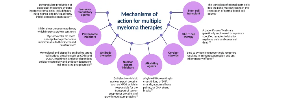
BCMA, B-cell maturation antigen; IL6, interleukin-6; MIP1α, macrophage inflammatory proteins; RANKL, receptor activator of nuclear factor-κB ligand; TNFα; tumor necrosis factor alpha; XPO1, exportin-1.
*Data from Tanenbaum, et al.9
Region-specific guidelines
References
Please indicate your level of agreement with the following statements:
The content was clear and easy to understand
The content addressed the learning objectives
The content was relevant to my practice
I will change my clinical practice as a result of this content